Gallery

explore exciting images from SunJin Lab

Gallery

Image

Mouse paw in RC152

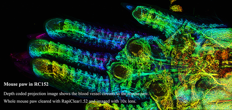
Depth coded projection image shows the blood vessel circuits in the mouse paw. Whole mouse paw cleared with RapiClear1.52 and imaged with 10x lens.
File size: 66,858 kB

Mouse liver in RC152

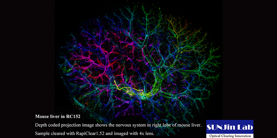
Depth coded projection image shows the nervous system in right lobe of mouse liver. Sample cleared with RapiClear1.52 and imaged with 4x lens. Courtesy of Mr. RYU NAKAMURA, System Development Section, NIKON Corporation File size: 29,702kB  

Mouse lymph node in RC152

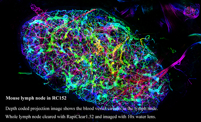
Depth coded projection image shows the blood vessel circuits in the lymph node. Whole lymph node cleared with RapiClear1.52 and imaged with 10x water lens.
File size: 44,975 kB

Mouse heart in RC152

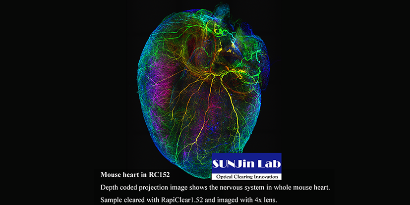
Depth coded projection image shows the nervous system in whole mouse heart. Sample cleared with RapiClear1.52 and imaged with 4x lens.
Courtesy of Mr. RYU NAKAMURA, System Development Section, NIKON Corporation
File size: 29,701 kB

Mouse spleen in RC152

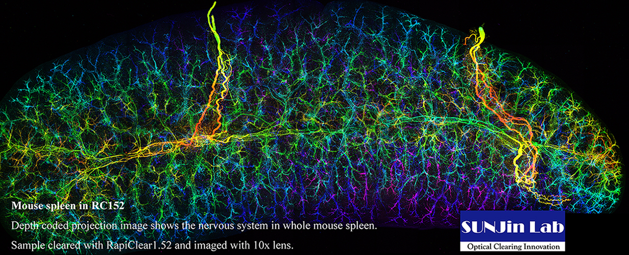
Depth coded projection image shows the nervous system in whole mouse spleen. Sample cleared with RapiClear1.52 and imaged with 10x lens. File size: 29,285 kB

Mouse kidney in RC152

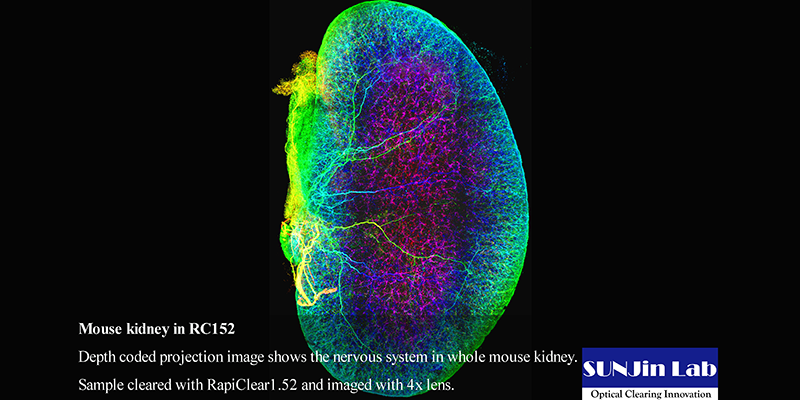
Depth coded projection image shows the nervous system in whole mouse kidney.
Sample cleared with RapiClear1.52 and imaged with 4x lens.
Courtesy of Mr. RYU NAKAMURA, System Development Section, NIKON Corporation
File size: 29,705 kB

Mouse brain in RC147

Depth coded projection image shows the Thy1-GFP mouse brain neuronal circuitry cleared with CLARITY-RapiClear 1.47 reagent.
File size: 25,153 kB

Mouse embryo in RC147

Depth coded projection image shows the E13.5 mouse embryo neuronal circuitry labeled with anti-TUJ1 antibody and cleared with CLARITY- RapiClear 1.47 reagent.
File size: 30,396 kB

Mouse cerebellum in RC152

Depth coded projection images show YFP-expressing cerebellum neurons in 550um mouse brain slices. Sample cleared with RapiClear1.52 and imaged with 25x oil lens.
File size: 17,083 kB

Mouse spinal cord in RC149

Depth coded projection images show YFP-expressing spinal cord neurons in 550um mouse brain slices. Sample cleared with RapiClear1.49 and imaged with 25x oil lens.
File size: 11,803kB

Drosophila brain in RC147

3D visualization of a single Drosophila optical lobe projection neuron.  Neuron arborization is labeled by mCD8::GFP (green), pre-synaptic terminals are labeled by anti synaptotagmin::HA Ab (magenta).
File size: 1,821 kB

Mouse hippocampus in RC149

Depth coded projection images show YFP-expressing hippocampal neurons in 550um mouse brain slices. Sample cleared with RapiClear1.49 and imaged with 25x oil lens.
File size: 14,494 kB

Mouse hippocampus in RC152

Depth coded projection images show YFP-expressing hippocampal neurons in 550um mouse brain slices. Sample cleared with RapiClear1.52 and imaged with 63x oil lens.
File size: 11,377 kB

Mouse brain in RCCS

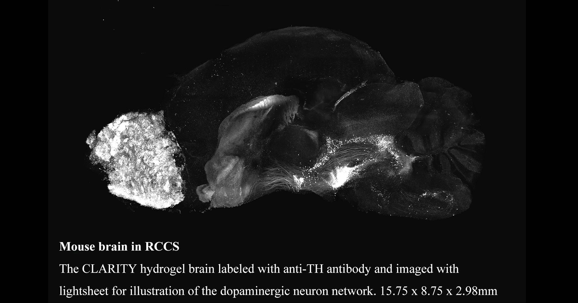
The CLARITY hydrogel brain labeled with anti-TH antibody and imaged with lightsheet for illustration of the dopaminergic neuron network. Volume Size: 15.75 x 8.75 x 2.98mm. Sample cleared with RapiClear CS and imaged with 5x lens.
File size: 1,679 kB

Mouse pancreas in RC152

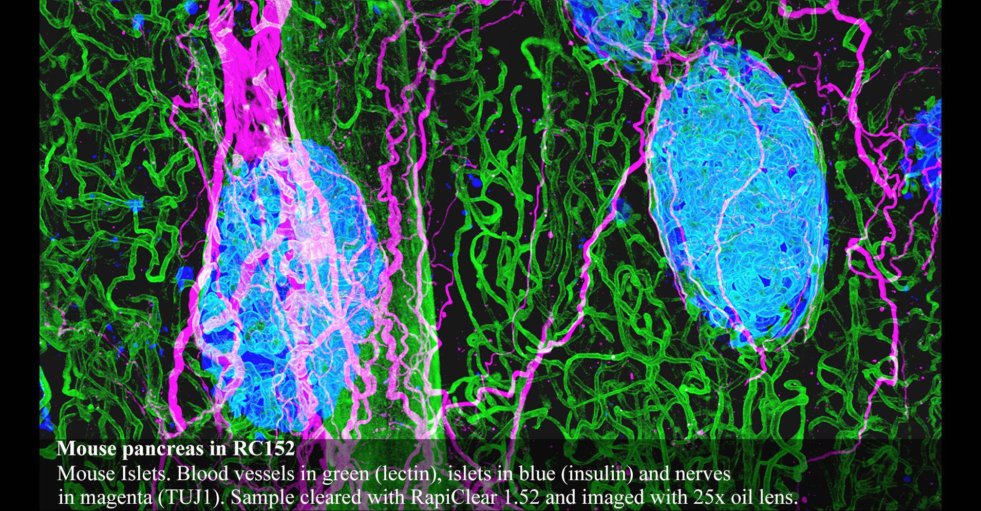
Mouse Islets. Blood vessels in green (lectin), islets in blue (insulin) and nerves in magenta (TUJ1). Sample cleared with RapiClear 1.52 and imaged with 25x oil lens.
File size: 5,316 kB

Mouse spinal cord in RC149

Depth coded projection image shows YFP-expressing spinal cord neurons in 550um mouse brain slices. Sample cleared with RapiClear1.49 and imaged with 25x oil lens.
File size: 33,948 kB

Mouse brain in RC152

Depth coded projection image shows YFP-expressing hippocampal neurons in 550um mouse brain slices. Sample cleared with RapiClear1.52 and imaged with SRRF super-resolution function in Andor Dragonfly confocal.
File size: 9,909 kB